
Grafting Strategy
The order of grafting is very important and should be discussed ahead of time with the surgeon. The grafts to the anterior portion of the heart are much easier to do and revascularization of the region of the LAD may be preferable before any lifting or turning of the heart is necessary.
It is important to maintain a high perfusion pressure until the graft is open. Try not to wait until the heart is displaced to increase the systemic pressure. It is much easier to start with a higher pressure and have it reduced back to normal by displacement of the heart, than to recover from hypotension while the heart is ischemic.33
Also relevant to the hemodynamic response is the severity of the coronary artery's stenosis. Mild stenosis tends to produce significant changes when the coronary artery is occluded suddenly; with a very severe stenosis, it is likely that collateral circulation has developed and the effect of occlusion is somewhat moderated.
Monitoring Limitations
There are unique challenges with monitoring during beating heart surgery due to displacement of the heart.
 |
| Figure 6. Beating Heart Surgery Monitoring Limitations |
- EKG, particularly
ST segment changes:
- The EKG as a tool becomes of limited use because of displacement of the heart. This alters the appearance of the EKG, which often decreases markedly in amplitude. In this situation, the ST segment changes may be falsely minimized, when in fact they represent 50% of the EKG complex (see figure 7).
- Pulmonary artery catheter:
- An abrupt elevation in pulmonary artery pressure or PA mean pressure is used as an indicator that the ventricular compliance has decreased or that mitral regurgitation has developed.
- Watch closely the CVP and PA pressure as they may be markedly elevated due to heart displacement, but volume in the right heart and pressure in the pulmonary vasculature may actually be low.
- Echocardiogram:
- Systolic thickening decreases or stops with ischemia and these regional wall motion abnormalities (RWMA) can be observed and followed on the echocardiogram.
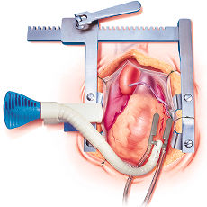
- The quality of the TEE image may deteriorate markedly due to obstruction of the view caused by pads placed behind the heart to bring it closer to the surgeon.
- Direct visual observation
of the heart itself:
- The affected area stops contracting and may be visible on the surgical field.
- Direct view of the heart in the surgical field is limited by the presence of the surgeon's hands
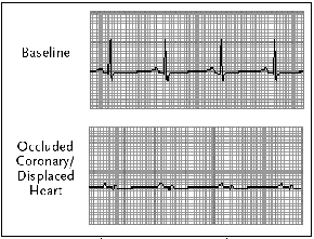 |
| Figure 7. Beating Heart Surgery EKG Changes |
|
|
||||||||||