|
STABILIZATION |
|
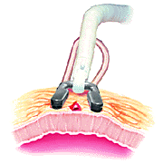
Target area stabilization is important for achieving a precise anastomosis on a beating heart. Suction Suction technology (e.g., Octopus¨2 Tissue Stabilization System) provides secure attachment of the stabilizers to the epicardium. Lifting and separating the stabilizer heads pulls tissue taut to isolate and immobilize the target area (Figure 6). Optimal stabilization is provided with motion reduction in all three planes (X, Y, Z).11,17
|
 Figure 6. The Octopus®2 Tissue Stabilizer immobilizes the target area by lifting the epicardium and pulling the tissue taut. |
|
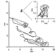
The motion reduction capabilities of the Octopus Tissue Stabilization System are depicted in Figure 7.
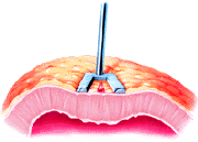
Compression Compression stabilizers use downward force to compress the myocardium and restrict its motion (Figure 8). In fact, cardiac output has been shown to be temporarily decreased as a result of direct ventricular compression with reduced stroke volume.19
|
|


