

|
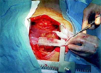
OM - PDA Exposure Techniques by A pericardial-based retraction system effectively supports the heart while allowing access to coronary arteries on the inferior and lateral walls of the heart (Figure 5). A mechanical stabilizer can then be used to immobilize the target area. The retraction system is composed of stockinet material (2cm x 30cm) and a 36-inch braided suture. To set up the retraction system: 1. Expose the posterior pericardium by manually retracting the heart to the patient's right. 2. Pass the suture through the pericardium at a point just behind the coronary sinus and directly beneath the origin of the most distal coronary artery (usually halfway between the left inferior pulmonary vein and the IVC). 3. Pass the suture through the midpoint of the stockinet. 4. Have an assistant gently retract the inferior portion of the heart. 5. Draw the stockinet into the pericardium and tie the suture. The middle of the stockinet is now attached to the posterior pericardium, and the two free ends of the stockinet are lying outside the chest. As traction is applied to the two free ends, the point of fixation moves in an anterior (not lateral) direction. The following techniques provide good vessel access:
To enhance exposure, rotate the table to the right and use the
Trendelenburg position. |

 |
|
|
||||||||||