|
CARDIAC MANIPULATION |
|
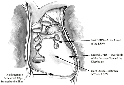
The first barrier to overcome with cardiac manipulation is fear of the unknown. Fear can be eliminated by trusting in the knowledge and experience of other beating heart surgeons. New skills, techniques, and tricks are required to manipulate a beating heart in order to obtain good coronary artery exposure while maintaining stable hemodynamics. Manipulation of a beating heart requires patience and slow, gentle maneuvers. Do not occlude coronary arteries until there is sufficient exposure and stabilization of the target area. The comfort level required to confidently perform the anastomosis, and indeed the success of the procedure, are directly related to good exposure and stabilization. The following procedures illustrate techniques for exposing the vessels on the posterior wall of the heart. For clarity, assume the use of a median sternotomy for access. Vessel Exposure Using Deep Pericardial Retraction Sutures Traction placed on deep pericardial retraction sutures (DPRSs) is used to rotate and vertically displace the apex of the heart. The importance of the DPRSs, used in combination with the Trendelenburg position and table rotation, for cardiac manipulation and vessel exposure cannot be overstated. However, traction placed on the DPRSs and pressure placed on the ventricle by sponges or other packing material can cause significant drops in blood pressure if they are not instituted gradually. DPRSs are placed in the following locations (Figure 2):
|
 Figure 2. Posterior pericardium with heart removed: Placement of DPRSs |


